
Preview









Description
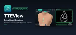
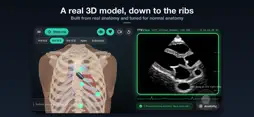
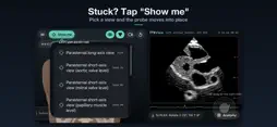
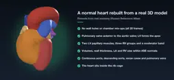
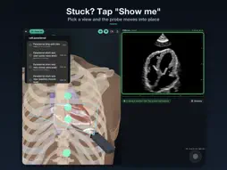
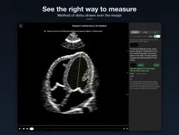
TTEView lets you practice the three steps of transthoracic echocardiography (TTE) on your iPhone, held sideways: finding the view, measuring, and grading. BUILT AND SUPERVISED BY A PRACTICING PHYSICIAN The scanning steps, the timing and position of each measurement, and the grading criteria are checked one by one against the guidelines and clinical practice. A REAL 3D MODEL, TUNED FOR NORMAL ANATOMY The normal heart was rebuilt for this app from real human anatomical data (Human Reference Atlas). Automatic checks cover the relations of the valves and chambers, papillary muscles and moderator band, continuous great vessels, and LV/RV volumes, wall thickness, LA size and RV diameters within ASE normal ranges, in all 20 frames of the beat. The heartbeat follows the order of electrical activation. Change how you hold the probe, and the echo image of that plane is generated on the spot. WHO IT IS FOR - Beginners who want to find the standard views themselves - Anyone practicing where and when to measure LVDd, Simpson's biplane, E/e' and TAPSE, through to guideline-based grading SCANNING - Choose an acoustic window on the body (left parasternal 3rd-5th intercostal spaces, apical, subcostal). Touch the right side of the screen to tilt the probe; swipe the left side to rotate it - The frame turns green when you hit one of 11 standard views (PLAX, PSAX at four levels, apical 4-, 5-, 2- and 3-chamber, subcostal 4-chamber, IVC) - When you are off, the app shows how far to rotate or tilt - The echo image is formed the way ultrasound images are (scattering, beam width, specular reflection). Color Doppler shows the flow through the valves MEASUREMENTS (27) - LV: IVSd, LVDd, PWd, LVDs, Simpson's method (A4C and A2C), M-mode LVIDd/LVIDs with FS and EF (Teichholz) - Diastolic function: E, A, DT, e' (septal and lateral), LA volume - Right heart: TAPSE, TR velocity, PR velocity (early and end-diastolic), IVC diameter (expiration and inspiration) - Hemodynamics: TR and PR gradients (4V²), RA pressure, PA systolic and diastolic pressure (≈ PCWP) and cardiac index, all the way to the Forrester class by echo - Aortic valve: LVOT diameter, LVOT VTI, AV Vmax, AV VTI - You place the M-mode line, the pulsed-wave Doppler gate and the continuous-wave line yourself. The waveform changes with your placement, and the app tells you whether the placement is right CASE PRACTICE - Scan hearts with myocardial infarction (wall-motion abnormalities by occlusion site, including RV infarction with proximal RCA occlusion) or valve disease (color Doppler of regurgitation and stenosis), then give your diagnosis - After you answer, an explanation shows which segments and views reveal the findings GRADING - Based on ASE/EACVI 2015 (chamber quantification), ASE 2025 (diastolic function), ASE 2010 (right heart), ESC/ERS 2022 (pulmonary hypertension) and ACC/AHA 2020 (valvular heart disease) - Reference values and sources (papers with DOIs) are available in the app WHAT MAKES IT DIFFERENT - You find each view with your own fingers (window, tilt, rotation) instead of jumping to it, with guidance on how far to move - Measurements are scored against the correct value on the same image - Anatomy mode (Pro): slice the real 3D model along the probe plane - No network, no ads, no account. One-time purchase, not a subscription FREE AND PRO - Free: scanning (grading, guidance and "Show me" for all 11 views), the LVDd measurement, and LAD infarct cases in Case Practice - Pro (one-time purchase): all 27 measurements, guideline-based grading, anatomy mode, all Case Practice cases and "Choose a case to view" PLEASE NOTE - The images and Doppler waveforms are simulated from a heart model. They are not images of real patients - In Case Practice, 2D, M-mode and Doppler measurements and grading are scored against the case heart - This app is for education. Do not use it for diagnostic or treatment decisions
What's New
Version 1.0.3 2026-11-16
- Case Practice now supports measurements: 2D, M-mode and Doppler (PW, CW and tissue Doppler) on the case heart, with grading (diastolic function, pulmonary hyp...- Case Practice now supports measurements: 2D, M-mode and Doppler (PW, CW and tissue Doppler) on the case heart, with grading (diastolic function, pulmonary hypertension, echo-based Forrester class and more). - Valve disease cases now show chronic changes: chamber dilatation, septal flattening, a dilated IVC, and LV wall thickening in aortic stenosis. - In myocardial infarction cases, mitral inflow and tissue Doppler now change with infarct size and left atrial pressure. - More realistic color Doppler: regurgitant and stenotic jets change shape from frame to frame, turbulence appears as a green mosaic, and a velocity color bar has been added. Technical Analysis
Package Overview Basic metadata
Bundle ID
com.tomoaki.tteview Current Version 1.0.3
Minimum iOS 17.0
Package Size App Store Size
Package Metadata 0 / 0 version records
Detailed binary, privacy manifest, framework, and Info.plist results have not been indexed for this package yet. The verified App Store and archive metadata above remains available.
Known Version Records
0 version records with stored package metadata
Stored metadata does not guarantee a package is currently available to download.
| Details | Version / IDs | Version date | Action |
|---|